
hiPSC-derived kidney organoids supply pseudo-proximal tubule cells
3D self-organization and nephrogenesis commences at day 7, after harvesting the progenitor cell combination and pelleting onto transwell filters (Fig. 1a). It was established that the organoid comprises optimum share and situation of proximal tubule cells on day 22. EpCAM+ inhabitants (renal tubules) was recognized as superset of proximal tubule cells labeled by a brush border marker, Lotus Tetragonolobus Lectin (LTL), and extra particularly by the albumin transporter, megalin (Fig. 1b). According to the sooner estimates, the organoid roughly comprises a 20-30% inhabitants of proximal tubule cells19. A 3D confocal reconstruction of the organoid is offered (Supplementary Movies 1 and a couple of).
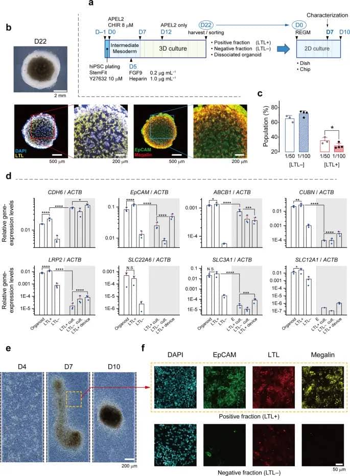
a The simplified protocol of hiPSC differentiation into intermediate mesoderm and formation of 3D kidney organoids, adopted by dissociation, sorting, and seeding the cells as sorted. Dissociated organoid, LTL + and LTL–, check with dissociated organoid cells, constructive fraction of MACS, and adverse fraction of MACS, respectively. b Select brightfield and confocal fluorescent photos (z-intensity projected) of kidney organoids on day 22. Immunochemistry is for EpCAM and proximal tubule markers, LTL and megalin. c Effect of LTL focus (dilution elements of 1/50 or 1/100) on the specificity of MACS, N = 4 experiments and the error bars signify commonplace deviation of information, *p < 0.05. d Relative gene expression ranges obtained from cells as sorted and cultured for 7 days in tradition dishes and PToC: Organoid, dissociated organoid cells on day 22; LTL+/–, constructive/adverse fraction of MACS merchandise. White space corresponds to samples obtained from as-dissociated (organoid) and as-sorted cells, whereas the grayed space signifies samples from cells cultured for 7 days. Error bars signify the usual deviation with N = 3 biologically unbiased experiments. N.S., not important for p > 0.05; *p ≤ 0.05; ** p ≤ 0.01; ***p ≤ 0.001; ****p ≤ 0.0001. e Evolution of LTL+ cells cultured on the membrane into aggregates, Scale bar, 200 μm. The yellow body exhibits the boundary between the cell combination and the monolayer. Scale bar, 200 μm. f Immunochemistry on day 7 for EpCAM, LTL, and megalin, markers of proximal tubules, in sorted cells extracted from kidney organoids and seeded on the PToC. For the LTL+ tissue fluorescent scans had been performed on choose elements on the proximity of aggregates as framed in (e). Scale bar, 50 μm.
On day 22 the organoids had been dissociated right into a cell suspension. The suspension was partially uncovered to LTL and subjected to MACS after the optimum antibody focus was decided (Fig. 1c). (Also see ‘Methods’ part and Supplementary Table 1 for particulars.) We additionally examined FACS for its larger diploma of specificity and characterised the merchandise as-sorted and cultured (Supplementary Fig. 1, 2 and Supplementary Data 1). However, finally we proceeded with MACS due to its functionality to supply cells with phenotypic options related sufficient to main proximal tubule cells (Supplementary Movies 3–6). Subsequently, the constructive and adverse fractions of the MACS merchandise had been collected and cultured for 7 days in 2D circumstances or lysed, as-sorted, together with remainder of the cell suspension. To decide if the proximal tubule compartment of the nephron was current in our organoids and study the impact of 2D tradition, we performed mRNA evaluation towards various related genes.

We discovered that the expression degree of CDH6 (Ok-cadherin), a selected marker of the proximal tubule progenitor cells16,26,27, elevated throughout tradition whereas the rise was extra pronounced for the adverse fraction (LTL–). Namely, the will increase had been 2.2-fold and 6.2-fold for the LTL+ and LTL– populations, respectively (Fig. 1d). In distinction, the 2D setting was not favorable for the remainder of markers examined, because the expression ranges dropped (ABCB1, CUBN, LRP2, SLC3A1, and presumably SLC12A1), or completely diminished (SLC22A6 (OAT1)).
The tissue cultivated from LTL+ cells cultured within the chips (Supplementary Fig. 3a,b) started to combination and kind tiny spheroid-like agglomerates on the membrane (Fig. 1e). Nevertheless, immunostaining confirmed the abundance of proximal tubule proteins in these cells. EpCAM, LTL, and megalin appeared with excessive distinction, solely in LTL+ cell aggregates that had been fashioned slowly and have become discernable round D7 (Fig. 1f). No aggregates had been noticed within the LTL– tissue layer (Supplementary Fig. 3c).
Introducing the LTL+/RPTEC coculture system
Following MACS, each LTL+ and LTL– fractions had been collected and cultured at 107 cells mL–1. In addition, a 50:50 coculture of every of the fractions with immortalized renal proximal tubule epithelial cells (RPTEC/TERT1 cells, hereafter known as RPTECs) had been examined (Fig. 2a). As proven within the shiny subject photos (Fig. 2b), LTL+ cells retrieved their organoid area of interest and started to combination. However, as soon as cocultured with RPTEC cells they flattened out, making it potential to check the barrier operate. It grew to become clear that LTL+ and RPTEC cells blended properly, supplied a confluent layer, and had been constructive to EpCAM (Fig. 2c). Whereas in distinction, though LTL– cells within the combination had been clustered they may not coalesce right into a steady tissue. The LTL+/RPTEC epithelial layer was hereafter known as the coculture. Confocal laser scans directed from the apical to the basal sides present the expression of apical (LTL, ZO-1, and Pgp) and basolateral (EpCAM) proximal tubule markers (Supplementary Movies 7–9).
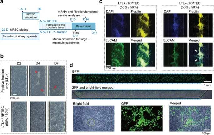
a Timeline (in days) demonstrating the coculture assay. hiPSCs are plated 23 days previous to the coculture as outlined earlier, adopted by RPTECs that are subcultured on day –5. Kidney organoids are harvested upon maturation, dissociated, and combined in equal parts with RPTECs. The cell combination is then launched into the chip. b Brightfield photos displaying the evolution of LTL+ and cocultured tissues with time. The constructive fraction-only cells begin to combination from ∼D4 and kind separable spheroids by D7 (as indicated by purple arrowheads), rendering the system impractical for filtration assessments. Whereas no detachment is noticed within the coculture with RPTECs. Scale bar, 200 μm. c Z-intensity projected fluorescent photos taken from immunostained samples on D7. DAPI, F-actin and EpCAM are represented in blue, yellow, and inexperienced, respectively. Once cocultured with RPTECs at a 50/50 ratio, LTL+ cells mix properly and make a confluent tissue layer. Coculturing the LTL– fraction with RPTECs nevertheless, doesn’t yield a confluent tissue layer. The tissue partially detaches as indicated by white arrowheads. EpCAM is faintly expressed all through this tissue and largely within the RPTECs. Gray dashed traces present the whereabouts of the PToC membrane. Scale bar, 200 μm. d Fluorescent, brightfield and merged photos of the cocultured tissue on D14. Top rows present that the channel is roofed by RPTECs and LTL+ cells equally in its entirety. High magnification photos on the backside row elucidate that the dissociated/sorted cells within the combination not solely kind small aggregates however are additionally distributed almost evenly all through the coculture tissue. Scale bars are in 1 mm and 100 μm on high and backside rows, respectively. Dashed traces are to information the attention.
For preliminary trials GFP-positive hiPSCs had been employed in cultivating the organoid with the intention to ease monitoring of the sorted cells. It grew to become clear that within the coculture tissue each cell varieties preserve their conformality over the complete layer. Organoid-derived cells that populate ∼50% of the epithelium are discernable (Fig. 2nd). By day 14 the aggregates had been assimilated into the coculture, though some clusters nonetheless remained. At this stage the tissue was deemed able to be characterised.
RPTEC and LTL+ synergy, improved mRNA ranges and filtration capability
Apparently, the comb border marker is expressed extra strongly in kidney organoid-derived LTL+ cells than within the RPTECs. Also, we noticed that within the coculture tissue LTL+ cells secrete their very own ECM within the proximity of RPTECs since each laminin and collagen IV seem densely the place LTL antibody is strongly expressed. Intense expression of Pgp within the coculture might recommend an improved capability within the elimination of exogenous substances. (Fig. 3a).
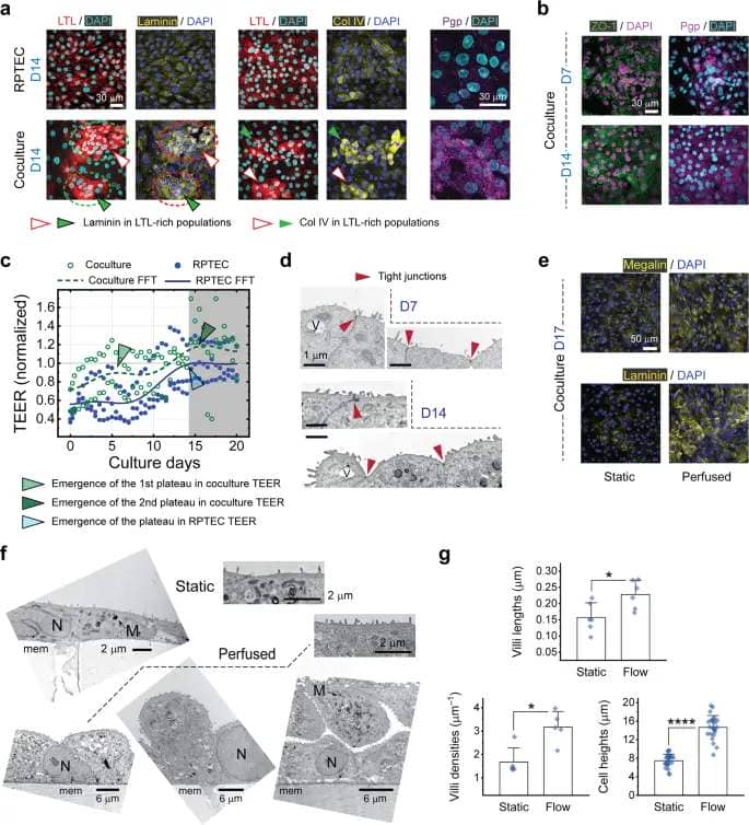
a Immunohistochemistry for LTL, laminin-111 (α1β1γ1) heterotrimer, collagen IV, and Pgp in single layer RPTEC and cocultured tissue layers on day 14, highlighting the enhancement of each ECM proteins within the latter. Laminin- and collagen-bound clusters, morphologically resembling islet-epithelial clusters in vivo, seem largely round LTL+ cells sorted off the organoids, as indicated by white and inexperienced arrowheads. Same applies for the proximal tubule apical protein, Pgp. Scale bars, 30 μm. b Appearance of tight junctions round LTL+ cell clusters on D7 and all through the complete coculture tissue on D14. In (a) and (b), image pares are taken from the identical pattern and scale bars are 30 μm. c Evolution of the transepithelial electrical resistance (TEER) with tradition time for the RPTEC-only and coculture tissue layers. In the coculture system the resistance will increase extra steeply and reaches the primary plateau of two (mild inexperienced arrowhead), presumably on account of smaller LTL+ cells within the combination. The second plateau (darkish inexperienced arrowhead) occurs at almost the identical time the resistance saturates within the RPTEC-only system, and it corresponds to the contribution of RPTECs within the combination (mild blue arrowhead). TEER is normalized to the ultimate steady-state worth of the RPTEC-only tissue. N = 3 and a couple of unbiased experiments for RPTEC-only and coculture instances, respectively. d TEM micrographs highlighting emergence and institution of tight junctions (darkish purple arrowheads) within the coculture monolayers on D7 and D14. Scale bar, 1 μm. V, vacuole. e Immunocytochemistry for megalin and laminin-111 exhibits enchancment in expression depth and distribution of each proteins within the coculture attributable to flow-induced shear stress. Scale bar, 50 μm (f) TEM micrographs of consultant cells from the coculture system examine static tradition and perfusion tradition circumstances. Formation of assorted epithelial tissue morphologies reminiscent of flat monolayers (that cowl almost the complete space of the membrane), monolayers with cell protrusions, and entrance to entrance (apical-apical) cell stacks is revealed within the latter. All the TEM micrographs are from the cells fixated on D14. Scale bars signify 2 μm and 6 μm for micrographs representing static (high) and perfused (backside) circumstances, respectively. M, mitochondria; N, nucleus; mem, PET membrane. g Quantification of the microvilli size/density and cell peak within the coculture system on D14. We outline the villi density because the depend of protrusions divided by the size of the cell membrane cross-section periphery, measured from particular person snapshots. Flow induced shear stress manifested development of not solely longer and denser microvilli but additionally taller cells evidencing for a squamous to cuboidal morphology translation. All samples are on D14. For quantification functions, from a complete of N = 3 unbiased TEM experiments per situation, n = 6, 5, and 24 cells had been randomly chosen to measure villi lengths, villi densities, and cell heights, respectively. Error bars signify the usual deviation of information.
The synergistic impact of mixing LTL+ cells with RPTECs can be evidenced by robust look of the tight junction protein. ZO-1 seems early on D7 within the coculture (Fig. 3b), corresponding with the primary plateau noticed within the TEER profile of Fig. 3c. In these plots stable and dashed traces signify smoothed estimation of the TEER developments for the RPTEC and coculture instances, respectively. The first plateau happens at an earlier date within the coculture (as proven by a lightweight inexperienced arrowhead), indicating a shorter contact inhibition interval. LTL+ cells are smaller in common and exhibit a steeper log part. The resistances converge round day 14, with the coculture (darkish inexperienced arrowhead) being barely extra resistive that the RPTEC tissue (mild blue arrowhead). Tight junctions are visualized within the TEM photos as properly (Fig. 3d). Our TEER measurement technique and setup have been reported earlier28,29. Bright filed photos present the evolution of tissue tradition layers on the TEER chips with time (Supplementary Fig. 3d). Clear barrier operate of the epithelial layer was verified previous to conducting transport measurements on D14 (Supplementary Fig. 3e).
To exhibit and quantify potential results of mechanical stimulus we uncovered the apical aspect of the tissue to flow-induced shear stress. The impact of media move on the tissue tradition is twofold. As against stagnant situation, move not solely helps to replenish essential elements of the media, e.g. development elements and hormones but additionally exerts shear stress on the underlying tissue. These two results have been not often distinguished within the literature of renal microphysiological methods. Here we circulated the tradition media on the static tradition situation as properly, however with a miniscule move fee (1 μL min–1) to make sure that the shear stress could be negligible whereas sustaining the availability of media elements. In this way we may distinctly discern and examine the impact of mechanical stimulus on the apical aspect of the endothelium. Proximal tubule cells are identified to reply to shear stimulus inside a sure vary. The steady shear stress alongside the proximal tubule is measured to differ from 0.1 to greater than 1 dyne cm–2 in vivo30,31. However, because of the tubuloglomerular suggestions the instantaneous move into the tubule is perhaps pulsatile quite than clean32. It is predicted that at a given fee a pulsatile move could be extra stimulating.
Previously Long et al. noticed that cell differentiation, proliferation, and to sure extent endocytosis had been enhanced at decrease shear stress ranges33. In the perfused media situation, we circulated the medium at a fee of 10 μL min–1 ensuing a median shear stress of 0.06 dyne cm–2 which is concerning the minimal reported values in vitro. As a outcome, we may infer that the instantaneously pulsating nature of the peristaltic pump (Supplementary Fig. 4a, b) might play a task within the enhancements in mobile morphology and performance noticed herein. Immunostaining revealed even such a small quantity of shear stress remarkably impacts the depth and distribution of megalin within the mature epithelia of the coculture and non-iPSC bilayer methods.
Various morphological and useful elements had been improved. Megalin, a cotransporter of albumin, was redistributed within the cytosol and it’s evident that laminin secretion has been improved (Fig. 3e). Electron microscopy additionally revealed important alterations in morphology of cells. Aside from the attribute options of mature proximal tubule cells with brush border constructions, vacuole- and mitochondria-rich areas had been noticed within the cocultured cells. Cells uncovered to flowing medium for under 3 days underwent apical and basal floor discount that’s indicative of squamous to cuboidal phenotype change (Fig. 3f)34,35. We had been additionally capable of quantify a few of these geometrical adjustments by analyzing numerous TEM photos. As illustrated within the graphs of Fig. 3g, cocultured cells grew taller and developed longer and denser microvilli. For such quantification functions we solely counted the cells within the monolayer and disregarded the small fractions of the tissue containing stratified cells.
Compared to both cell supply, the mRNA expression ranges of sure key genes had been elevated within the cocultured epithelium (Fig. 4a). However, discrepancies had been noticed between these ranges and the protein expression ranges of the corresponding markers, as revealed by immunostaining. For instance, though ABCB1 was expressed at almost the identical degree within the RPTEC and coculture tissue samples, Pgp was extra abundantly discovered within the latter. The glucose and albumin transporters, SGLT2, and LRP2, respectively, had been each elevated in mRNA and protein ranges. LRP2 was surprisingly elevated greater than 100-fold.
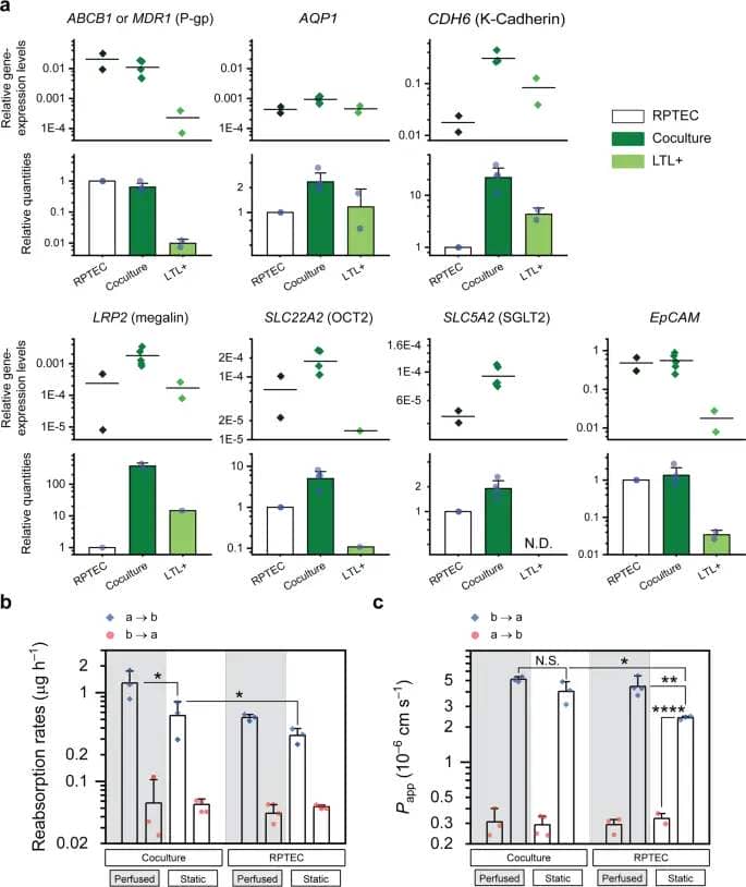
a Expression ranges of sure genes within the RPTEC/LTL+ coculture present enchancment over these of every element. Functional, ABCB1 (MDR1), AQP1, LRP2, SLC22A2 (OCT2), SLC5A2 (SGLT2), and structural, CDH6 (Ok-Cadherin) and EpCAM, relative gene expression ranges (versus these of the ACTB degree), are accompanied by relative portions (normalized to the values of the RPTEC tissue). Except for ABCB1 and EpCAM, the expression ranges of different genes had been considerably elevated within the cocultured tissue. There was no detrimental impact on mRNA expression ranges; N = 2 and 5 biologically unbiased experiments for RPTEC/LTL+ monocultures and the coculture case, respectively; Error bars signify the usual deviation of information. Reabsorption charges of glucose (b) and obvious permeabilities (Papp) to Rh123 (c) measured on D14 for each RPTEC-only and cocultured tissue layers subjected to static and perfused tradition circumstances. Vectorial transport is verified in all instances. Transfer charges had been estimated by conducting linear regression on time course knowledge. a → b, apical to basal; b → a, basal to apical; Measurements taken from a minimal of N = 3 unbiased chips; Error bars signify SD of the imply; N.S., not important for p > 0.05; *p ≤ 0.05; **p ≤ 0.01; ***p ≤ 0.001; ****p ≤ 0.0001.
We hypothesized such enhance within the transporter protein ranges may result in improved energetic transepithelial transport. To check our speculation, we quantified the reabsorption fee of glucose and the excretion fee of rhodamine 123 (Rh123), as measures of SGLT2 and Pgp exercise, respectively. We confirmed that the apical to basal (a → b) transport charges of glucose within the coculture had been improved by about 1.75 and a couple of.50 instances over that of the RPTEC-only monolayer in static and perfused tradition circumstances, respectively (Fig. 4b). Among all 4 circumstances examined, whereas a → b switch charges of the substrate had been considerably larger than these of the reverse (b → a) route, there have been no important adjustments within the reverse switch charges. Both observations affirm the vectorial nature of glucose transport.
Apparent permeabilities (Papp) to the Pgp substrate, Rh123, are proven in Fig. 4c. Since the excretion charges (b → a) are larger than the absorption charges (a → b), the transport is unidirectional in all 4 instances examined. In addition, the efflux ratio of the substrate, outlined because the ratio of excretion to absorption charges, has been elevated by 90% within the coculture system underneath static tradition situation, confirming substantial enchancment in xenobiotic deposition capability.
Establishing and characterizing the RPTEC/HUVEC bilayer chip alongside
To create utterly remoted proximal tubule epithelial and peritubular endothelial channels we tailored our protocol to make sure a completely sealed RPTEC tissue is fashioned earlier than introduction of HUVECs (Fig. 5a). This not solely prevented intermix of EGM2 and REGM that may have in any other case triggered detachment of HUVECs (Supplementary Fig. 4c, d) but additionally allowed us to check the consequences of endothelial cells on a maturated proximal tubule epithelial tissue. Prior to seeding we coated the membrane at either side solely with a cell adhesion agent and allowed the cells to secrete their very own ECM. While no exterior ECM protein was utilized, the bilayer was safely maintained for at the very least 10 days after which HUVECs started to detach (Supplementary Fig. 4e and Supplementary Movies 10 and 11).
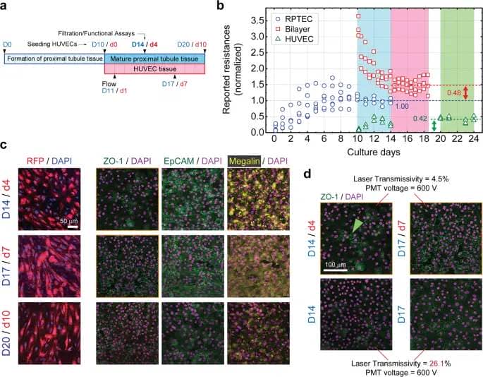
a The timetable of generic cell seeding and upkeep processes. HUVECs aren’t included for the single-layer gadgets. b Time course of reported resistances of the RPTEC-only (blue circles), bilayer (purple squares), and HUVEC-only (inexperienced triangles) tissue layers. For the bilayer gadgets as soon as HUVECs are launched on D10, the general resistance jumps upon formation of a confluent endothelial layer after which begins to say no till reaching regular state on D14/d4. Dashed traces point out the typical resistances obtained over the past 4 days of tradition (coloured bars) for every case. All the measured values are normalized to that of the RPTEC TEER at regular state, i.e. 60 Ω cm2 (blue dashed line). Green and purple two-headed arrows point out, respectively, the reported resistance of the HUVEC-only layer and the resistance increment measured upon addition of HUVECs to the RPTEC-only tissue. A steady bilayer is fashioned on D14/d4. RPTECs had been cultured on N = 6 gadgets, out of which N = 3 had been rendered bilayer upon addition of HUVECs on D10. N = 3 gadgets had been devoted for the HUVEC-only tissue. c Immunohistochemistry for ZO-1, EpCAM, and megalin in RPTEC tissue of the bilayer system together with fluorescent photos of corresponding RFP-tagged HUVECs, displaying the evolution of tight junctions, diploma of re-epithelialization, and distribution of the albumin transporter, respectively. Tissue exercise and integrity is peaked between D14/d4 and D17/d7. Scale bar, 50 μm. d Effect of HUVECs on the depth of ZO-1 expression; In the presence of HUVECs tight junctions appeared from D14/d4 (inexperienced arrowhead) onward and have become clearly seen on D17/d7, whereas within the absence of HUVECs they appeared faintly, even at a significantly larger confocal laser transmissivity. No Triton X was used on this experiment. Scale bars are 100 μm. All fluorescent photos are confocal z-intensity projected. Scale bar, 100 μm.
Similar to the single-layer coculture system, we probed the general electrical resistance of the RPTEC/HUVEC system throughout the complete course of tradition in intervals. Devices with single-layer RPTEC and HUVEC tissue had been used for comparability and all values had been scaled with respect to the typical RPTEC TEER measured throughout final 4 days of tradition, that’s 60 Ω cm2 (Fig. 5b). The total resistance overshoots throughout formation of a confluent HUVEC monolayer from D10/d0 to D13/d3 after which plateaus off previous d4. However, the amplitude of resistance bounce upon creation of the bilayer, as indicated by the purple arrow in Fig. 5b, is just not considerably totally different from the resistance of the HUVEC-only tissue (inexperienced arrow). As a outcome, although the bilayer resistance profile appears steady in the course of the tradition, it’s unlikely that introducing HUVECs has considerably affected the transepithelial resistance of the RPTEC layer. Usually, RPTEC monolayer cultures exhibit TEERs round 0.1 to 1 kΩ cm2 36,37, whereas the proximal tubule tissue in vivo is thought to show values which can be an order of magnitude smaller. It is commonly argued that the leakier barrier operate of the tissue in vivo is because of a better transcellular or paracellular transport38. Apparently, and presumably for a similar motive, the resistance values of some ten Ω cm2 measured herein lie between these of the beforehand reported values for RPTEC monolayers and the proximal tubule tissue in vivo. Later we set up that HUVECs certainly play a task in such enchancment.
It is properly established {that a} paracrine signaling community is fashioned as soon as RPTECs are cocultured with endothelial cells, resulting in improved RPTEC proliferation fee and differentiation39. In our case, proof of interplay and such signaling between HUVECs and RPTECs was discernable from d4 of the bilayer formation. We noticed that within the presence of HUVECs, epithelial tissue integrity and presumably operate each peak someday between days 4 and seven of the bilayer formation as evidenced by unwavering look of EpCAM/ZO-1, and expression of maximally dispersed (evenly distributed) megalin on d7 (Fig. 5c). Since megalin is a cellular albumin transporter an improved cytosolic supply of albumin, already current in tradition medium, is concluded.
This is noticeable as a result of in distinction to some earlier reviews the place each cell varieties had been introduced collectively at almost the identical time6, we observe enchancment of proximal tubule tissue integrity and performance even after 10 d of tradition. As noticed by cross-sectional transmission electron microscopy (TEM), 3 μm-wide pores of the PET membrane had been the one potential channels by way of which these two cell varieties may talk (Supplementary Fig. 3b). Indeed, a porosity of ∼5% was sufficient to ascertain enough cell-cell contact. In addition, the expression degree of RPTEC tight junction protein has been distinctly elevated within the bilayer system (Fig. 5d) just like an earlier report6, and so is its look frequency. As a outcome, all of the characterizations and useful assessments had been centered on D14 of the epithelial layer tradition whatever the cell supply. Notice that ZO-1 was expressed vividly within the coculture tissue on D14, whereas it has faintly appeared within the RPTEC-only single layer system on the similar tradition situation. The RPTEC/HUVEC bilayer system was subjected to an analogous diploma of scrutiny because the coculture tissue.
Protein profiling make clear morphological enhancements in proximal tubule cells
We estimated the spatiotemporal expression patterns of sure proximal tubule-specific proteins by conducting intensive immunofluorescence observations. Briefly, fluorescent intensities representing the expression ranges of a number of antibodies had been quantified by analyzing stacks of confocal photos taken throughout the cell-laden membranes. The process is illustrated utilizing consultant cross-sectional fluorescent photos of the RPTEC/HUVEC bilayer and the corresponding depth profiles (Fig. 6a, b). Δd denotes the gap between the positions of the height expression degree of every antibody and the height expression degree of DAPI, as a measure of nuclei whereabouts. By plotting values of Δd towards tradition days we may hint the motility of the proteins within the epithelial layer.
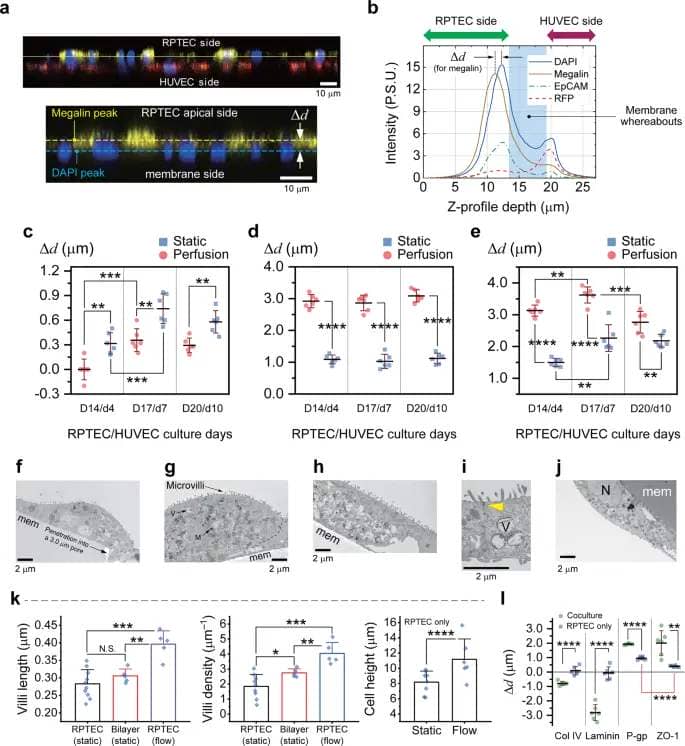
a Select cross sectional fluorescent confocal picture of the RPTEC/HUVEC bilayer system, immunostained for megalin, illustrating the definition of the relative distance of the protein of curiosity (e.g. megalin) measured from the middle of nuclei. Scale bars are 10 μm. b Representative z-intensity profiles of fluorescent alerts obtained by averaging emission intensities of assorted markers all through a laser scan space of 0.1 cm2 with a Δz (pitch) of 200 nm. Average proximal tubule-specific marker-to-nuclei distance in RPTECs (bilayer) obtained underneath static and perfused tradition circumstances for (c) megalin, (d) LTL, and (e) SGLT2; N = 2 unbiased chips had been used to make n = 3 random measurements from every; Error bars point out commonplace deviation. Statistically important variations between knowledge pairs are indicated by asterisks, *, **, ***, ****, for p ≤ 0.05, 0.01, 0.001, and 0.0001, respectively. Select cross-sectional TEM photos spotlight the looks of the apical membrane of RPTECs in numerous tradition circumstances on D14/d4: (f) single layer underneath static, (g) bilayer underneath static, and (h) single layer underneath perfusion tradition circumstances. The close-up TEM of (i) reveals a good junction (yellow arrowhead) fashioned between two adjoining RPTECs underneath perfusion tradition. j A HUVEC on the reverse aspect of the membrane. Scale bars are 2 μm. All the micrographs are from the cells fixated on D14/d4. M, mitochondria; N, nucleus; V, vacuole; mem, PET membrane, arrows level to tight junctions, and the dashed traces present cell-cell boundaries. okay Quantification of the microvilli size/density and the RPTEC peak. We outline the villi density because the depend of protrusions divided by the size of the cell membrane cross-section periphery, measured from particular person snapshots. RPTECs/HUVECs are on D14/d4. Clearly RPTECs within the proximity of HUVECs developed denser apical microvilli, ensuing a better floor space. In addition, the microvilli density distribution is narrower within the bilayer configuration. No important distinction in villi lengths was noticed between the one layer and bilayer instances in static tradition situation. Flow induced shear stress manifested not solely longer but additionally a bigger density of microvilli. For quantification functions, from a complete of N = 3 unbiased TEM observations per situation, n = 9, 5, and 5 RPTEC micrographs had been randomly chosen from single layer (static), bilayer (static), and single layer (move) tradition circumstances, respectively, to measure villi lengths and densities. To measure cell heights, n = 8 and n = 9 micrographs had been examined for static and move circumstances, respectively. Error bars signify the usual deviation of information. l Average distances of basement and apical proteins from the nucleus illustrated for RPTEC-only and coculture tissues developed underneath move tradition situation. Data obtained by z-intensity profiling of fluorescent photos. Statistics are derived similarly to (c–e).
For the case of megalin, the farther it seems from the nuclei the upper likelihood of it being sure to the apical membrane, i.e., stay at an inactive state. We observed that within the bilayer system if HUVECs are seeded first, megalin which is initially expressed on the apical membrane, turns into internalized throughout 7 days of RPTEC tradition (Supplementary Fig. 5a). Whereas, when RPTECs are seeded first, it will be utterly taken into the intracellular milieu on D14, suggesting a peak endocytosis exercise (Fig. 6c and Supplementary Fig. 5b)40. Data offered herein and the fluorescent photos of Fig. 3e clarify that perfusion not solely will increase the exercise of megalin but additionally its abundance within the cytosol. Such enchancment is mirrored in a better reabsorption fee of albumin. In line with earlier reviews of cell peak/compactness enhance underneath fluid shear stress31, by protein profiling right here we discovered that the apical markers, LTL and SGLT2, seem extra distant from the nucleus (Fig. 6d, e).
Cross-sectional electron microscopy of the cells on the highest layer at day 14 revealed the distinctive results of shear stress and proximity with HUVECs on the non-iPSC epithelia as properly (Fig. 6f–j). Self-secreted ECM elements subtle by way of the membrane pores offering a communication path with HUVECs. Even although the cells had been squamous as compared with 3D tubular constructions5,7, tight junctions had been clearly discernable. Observation of tight junctions confirms the impervious nature of each RPTEC-only and RPTEC/LTL+ coculture epithelia and is important to validate our transcellular transport assessments. Tight junctions within the RPTEC-only tissue started to kind at a later stage than these within the coculture monolayer system. It additionally seems that they turn into tighter underneath perfusion tradition, though this has not been statistically confirmed (Supplementary Fig. 6).
Regardless of the epithelial cell supply, the density and size of villi in addition to the cell peak, all elevated underneath the affect of shear stress, resembling the situation in vivo. Interestingly, even within the absence of mechanical stimuli denser villi appeared on RPTECs in touch with HUVECs (Fig. 6k). Larger and denser inhabitants of villi manifest in a extra environment friendly endocytosis and transcytosis.
On D14 we measured the typical relative distances of basement (laminin & collagen IV), subapical (ZO-1), and apical (Pgp) proteins from the nucleus within the single layer RPTEC-only and coculture methods (Fig. 6l). Since each the ECM and the apical proteins are expressed farther from the nuclei within the cocultured tissue, it’s gathered that the proximal tubule cells of the coculture tissue grew taller in common. Undoubtedly, RPTEC tissue has a extra uniform thickness throughout the membrane and the collective diploma of cell orientation and polarization is larger (smaller error bars for Pgp and ZO-1). Also discover that ZO-1 is expressed clearly beneath the apical Pgp within the RPTEC-only tissue. Quantification of cell heights primarily based on TEM observations of particular person cells didn’t fairly result in the identical outcomes as these offered in Fig. 6l; nevertheless, the development was related. We consider the discrepancy roots within the technique of z-intensity profiling the place the typical relative distances measured between apical and basal markers are shortened as a result of the PET membrane is just not flat. In addition, since apical proteins aren’t essentially expressed proper on the height of the cells measuring cell heights from the membrane to the cell apex would lead to bigger values. Having stated that the impact of shear stress on cell peak was extra pronounced within the coculture, i.e. 37% enhance within the RPTEC solely tissue versus 99% within the coculture (cell heights in Fig. 3g and Fig. 6k).
Investigating the affect of shear stress and proximity to HUVECs on RPTEC mRNA ranges and filtration efficiency
The protocol for the formation of the RPTEC/HUVEC bilayer is as soon as once more depicted in Fig. 7a. To decide whether or not the presence of HUVECs would have an effect on markers of proximal tubule construction and performance, we performed qPCR evaluation. Apart from morphological enhancements noticed within the microvillar apical membrane, we observed mRNA ranges of sure RPTEC genes are elevated underneath move induced shear stress and within the presence of HUVECs. On the one hand AQP1, EpCAM, MDR1, OCT2, and presumably SGLT2 are upregulated within the mature bilayer system subjected to perfusion move, however neighborhood with HUVECs underneath media perfusion has triggered the expression ranges of AQP1, EpCAM, MDR1, and OCT2 to extend (Fig. 7b).
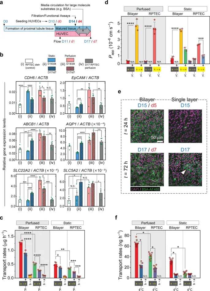
a The generic protocol for quantification of renal reabsorption and excretion utilizing the PToC. Measurements begin on day 14 no matter the assay in hand and the tissue layer configuration. The media in each epithelial and vasculature microchannels are circulated to enhance diffusion of large-molecule substrates (e.g. BSA-AF488). Transfer charges (reabsorption of albumin/glucose or obvious permeability to Rh123) had been estimated by conducting linear regression on time course knowledge. b Relative expression ranges (vs. ACTB) of structural genes, CDH6 (Ok-Cadherin) and EpCAM, and useful genes, specifically, ABCB1 (MDR1), AQP1, SLC22A2 (OCT2), SLC5A2 (SGLT2), vs. the extent of ACTB gene within the RPTEC-only and RPTEC/HUVEC methods. Four teams are offered, specifically, (i), RPTECs on D14 within the tradition dishes; (ii), RPTECs of the bilayer underneath static tradition situation; (iii), RPTECs of the bilayer underneath perfused tradition circumstances; (iv) Single layer RPTECs underneath perfused media. Proximity with HUVECs underneath media perfusion has augmented the expression ranges of AQP1, EpCAM, MDR1, OCT2, and SGLT2 (examine teams iii and iv). Independently, perfusion has elevated the expression ranges of the identical genes (with the potential exception of SGLT2) within the co-culture (examine teams ii and iii). n = 5 PCR replicates had been analyzed for every gene/pattern obtained from one set of experiment. c Transfer charges of the glucose probe, 2NBDG measured in static and perfused tradition circumstances. Both reabsorption (a → b) and reverse switch charges (b → a) had been quantified. The inhibitory impact of phlorizin (P.) on glucose transport was additionally examined; n = 3 unbiased chips. d Apparent permeabilities to Rh123 in each excretion (b → a) and reverse (a → b) instructions. Verapamil (V.), a contender for Rh123 extrusion, was utilized to attenuate Rh123 efflux and make sure the operate of Pgp; N = 3 unbiased chips. e Fluorescent confocal z-stacked photos of the RPTEC tissue layer in bilayer and single layer configurations. A significantly larger quantity of BSA was precipitated within the basolateral milieu of RPTECs within the bilayer system indicating a better consumption of the substrate. Some BSA is precipitated within the single layer RPTEC on D17 (white arrowhead). Scale bar is 50 μm. f Transport charges of AF488-conjugated bovine serum albumin (BSA-AF488) in each instructions. In this examine, we examined the impact of lowering the incubation temperature (to 4 °C) on selective transport of BSA-AF488; minimal N = 6 unbiased chips. In (b–d, f) two-sample t-test was run between pair of information units as indicated. Error bars signify commonplace deviation; N.D., not detected; N/A, not accessible; N.S., not important; *, **, ***, ****, for p ≤ 0.05, 0.01, 0.001, and 0.0001, respectively. a → b, apical to basal; b → a, basal to apical.
Thus, we anticipated that the epithelium delivered to the proximity of HUVECs might exhibit not solely a denser construction but additionally larger xenobiotic elimination and glucose uptake charges. These suppositions had been confirmed by in-depth assessments of glucose reabsorption and Rh123 excretion charges, indicating that the extent of proteins comparable to SGLT2 and MDR1 (SGLT2 and Pgp) had been enhanced as properly (Fig. 7c, d). The a → b glucose reabsorption charges had been considerably improved in presence of HUVECs and underneath perfusion move, independently, whereas the b → a charges had been almost unaltered. Also, phlorizin as a basic aggressive inhibitor of each SGLT1 and SGLT241,42,43, successfully blocked reabsorption of glucose.
Although LRP2 was not stably detected, presence and correct operate of megalin was confirmed in RPTECs by analyzing albumin reabsorption charges. We confirmed that the albumin uptake capability of RPTECs will increase significantly within the presence of HUVECs. This is evidenced by accumulation of BSA within the basolateral aspect of RPTECs within the bilayer system (Fig. 7e). In addition, the bilayer displayed a better fee of albumin a → b transcytosis (Fig. 7f). Lowering temperature in the course of the measurements appears to have significantly diminished reabsorption charges as investigated beforehand44,45.
These outcomes are fascinating, notably as a result of the reabsorption and excretion charges are elevated although, intuitively, the endothelial layer might hinder the switch of any biomolecule throughout the channels. Paracrine signaling between the 2 cell varieties has improved megalin, SGLT2, and Pgp operate to the extent that glucose, albumin, and Rh123 are all transferred at significantly larger charges throughout the bilayer tissue. In addition, apical microvilli have turn into denser within the RPTECs cocultured with HUVECs even within the absence of mechanical stimuli (Fig. 5k). Notice that within the RPTEC/HUVEC bilayer system there was no tangible enchancment within the total efflux ratio of Rh123, whereas the glucose and albumin reabsorption charges had been each elevated. Lastly, we evaluated the capability of the basolateral xenobiotic transporter, OCT2, within the single layer and bilayer methods. OCT2 is answerable for the uptake of chemotherapeutic medicine reminiscent of oxaliplatin and cisplatin which can be identified to trigger drug-induced kidney harm46. Using cimetidine as an OCT2 inhibitor we confirmed selective uptake of cationic fluorescent EtBr and vectorial uptake of 4-Di-1-ASP (ASP+), each generally known as OCT2 substrates (Supplementary Fig. 7). However, in distinction to albumin, glucose, and Rh123 filtration assays, the place HUVECs proved to enhance the transport charges within the bilayer, the RPTEC single layer was extra environment friendly in absorbing ASP+ because the substrate was blocked by HUVECs.
")